Improving Limit of Detection
by injecting increasing sample volumes
3.3.2.
Separation of Mouse IgG from bovine serum albumin on Protein A Sepharose microcolumn. Note broadening of peak of non-retained BSA, while eluted IgG yields narrow peaks of increasing amplitude. Estimated limit of detection is 1μg IgG in 25μL of injected sample.


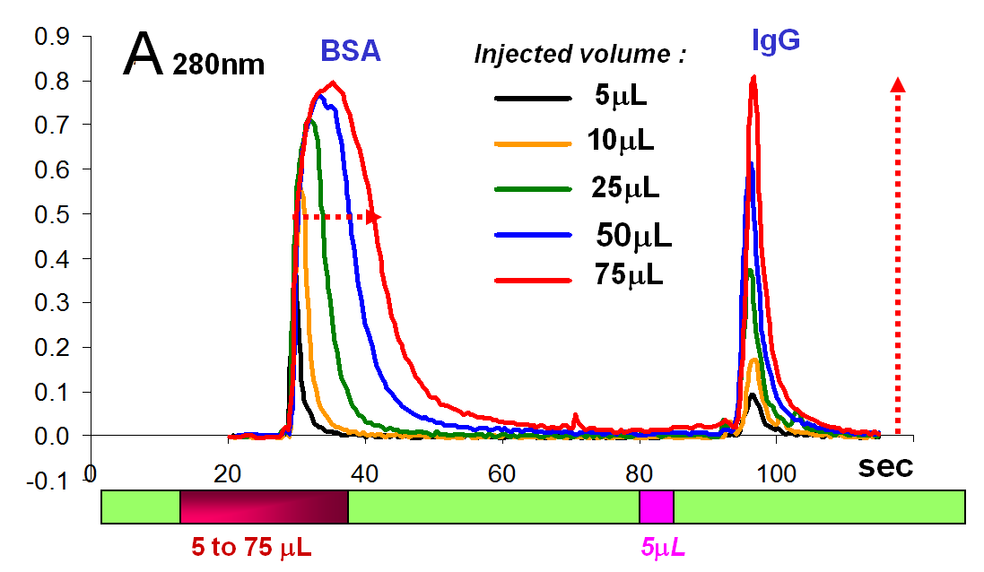
Sample: IgG 2μg/μL, BSA 4,6mg/mL. Mobile phase PBS (0.14MNaCl, 3.8mMKCl, EDTA 1mM, phosphate 18mM, pH=7.4.) Eluant 1M HCl, volume 5μL. Sample on column flow rate 5μL/sec, elution flow rate 2μL/sec. Column capacity 20mg/mL, column volume 10μL. Flow cell volume 6μL, optical path 3mm.